-
About University
- Mission & Brand Strategy
- University Leadership
- Rector's Welcome
- History
- Regulatory Documents
- Contacts
- Staff
- International Recruitment
- Partners
Applicants- Why Sechenov University
- Degree Programmes in English
- Preparatory Courses
- Non-Degree Programmes
- Transfer from other Institutions
19.01.2021Rare kidney cancer: identification by genetics
 The medics and scientists from Sechenov University performed a number of tests to diagnose an extremely rare type of renal cancer which is seen only in very few patients.
Genetic analysis plays an important role in the diagnosis of ially when doctors encounter a case with unusual manifestations, no known family history, or non-typical localisation of the tumour. Medics from Sechenov University and their colleagues from several other organisations teamed up to treat a patient who turned out to have kidney giant leiomyosarcoma — an extremely rare type of renal tumour. They used radiography, microscopy, immunohistochemistry, molecular genetic profiling, and chromosomal microarray analysis to substantiate the diagnosis. The case has been reported in the journal Frontiers in Oncology.
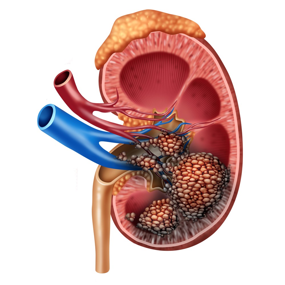
Kidney giant leiomyosarcomas account for only 0.12% of all renal malignancies and are usually found in females aged 50–60. In July 2014, a 67-year-old woman was admitted to a urology clinic at Sechenov University. She had an enlarged abdomen, dull pain in the lumbar region on the right, palpitation, and moderate weakness. Ultrasound examination revealed a 176 × 164 mm mass in the right kidney, and computed tomography showed that the organ was displaced upwards and practically replaced by a spherical tumour.
The medical team decided to perform nephrectomy of the right kidney. The resected tumour had a size of 300 × 253 × 150 mm and weighed 4 kg. Despite the successful surgery, the patient died in the post-operative period.
Microscopy studies of the tumour revealed plexiform bundles of spindle-shaped cells with large hyperchromatic nuclei, while immunohistochemistry showed positive reaction for smooth muscle actin and desmin, but negative reaction for CD99, CD43, CD117, S-100, MCK/PCK, and DOG-1. These findings point at Grade 3 Kidney Leiomyosarcoma.
Next generation sequencing identified no mutations associated with any genetic syndrome. The authors did not find single nucleotide variants (SNVs) in the genes usually mutated in this cancer — TP53, RB1, ATRX, and PTEN — as well as in the less commonly mutated genes ATM and EGFR2.
Chromosomal microarray analysis (CMA) revealed multiple chromosomal aberrations in the tumour. It contained a considerable number of gains and losses of chromosome fractions. The team discovered that 60 genes — including 2 oncogenes (GLI3, HMGA2), 6 tumour suppressor genes, and SP1 transcription factor gene — were truncated by chromoanasynthesis.
The authors of the study conclude that a thorough investigation of the genetic causes can serve as an important tool to keep track of rare cancer cases. Accumulation of such data will help understand the molecular aetiology and pathogenesis of tumours, resulting in better treatment and prognosis.
This case has been jointly investigated by Sechenov University, Research Centre for Medical Genetics (Moscow), Genomed Ltd. (Moscow), and Burdenko Main Military and Clinical Hospital (Moscow).
Read more:
Embed on website
Rare kidney cancer: identification by genetics
 The medics and scientists from Sechenov University performed a number of tests to diagnose an extremely rare type of renal cancer which is seen only in very few patients.
Genetic analysis plays an important role in the diagnosis of ially when doctors encounter a case with unusual manifestations, no known family history, or non-typical localisation of the tumour. Medics from Sechenov University and their colleagues from several other organisations teamed up to treat a patient who turned out to have kidney giant leiomyosarcoma — an extremely rare type of renal tumour. They used radiography, microscopy, immunohistochemistry, molecular genetic profiling, and chromosomal microarray analysis to substantiate the diagnosis. The case has been reported in the journal Frontiers in Oncology.
Kidney giant leiomyosarcomas account for only 0.12% of all renal malignancies and are usually found in females aged 50–60. In July 2014, a 67-year-old woman was admitted to a urology clinic at Sechenov University. She had an enlarged abdomen, dull pain in the lumbar region on the right, palpitation, and moderate weakness. Ultrasound examination revealed a 176 × 164 mm mass in the right kidney, and computed tomography showed that the organ was displaced upwards and practically replaced by a spherical tumour.
The medical team decided to perform nephrectomy of the right kidney. The resected tumour had a size of 300 × 253 × 150 mm and weighed 4 kg. Despite the successful surgery, the patient died in the post-operative period.
Microscopy studies of the tumour revealed plexiform bundles of spindle-shaped cells with large hyperchromatic nuclei, while immunohistochemistry showed positive reaction for smooth muscle actin and desmin, but negative reaction for CD99, CD43, CD117, S-100, MCK/PCK, and DOG-1. These findings point at Grade 3 Kidney Leiomyosarcoma.
Next generation sequencing identified no mutations associated with any genetic syndrome. The authors did not find single nucleotide variants (SNVs) in the genes usually mutated in this cancer — TP53, RB1, ATRX, and PTEN — as well as in the less commonly mutated genes ATM and EGFR2.
Chromosomal microarray analysis (CMA) revealed multiple chromosomal aberrations in the tumour. It contained a considerable number of gains and losses of chromosome fractions. The team discovered that 60 genes — including 2 oncogenes (GLI3, HMGA2), 6 tumour suppressor genes, and SP1 transcription factor gene — were truncated by chromoanasynthesis.
The authors of the study conclude that a thorough investigation of the genetic causes can serve as an important tool to keep track of rare cancer cases. Accumulation of such data will help understand the molecular aetiology and pathogenesis of tumours, resulting in better treatment and prognosis.
This case has been jointly investigated by Sechenov University, Research Centre for Medical Genetics (Moscow), Genomed Ltd. (Moscow), and Burdenko Main Military and Clinical Hospital (Moscow).
Read more:


